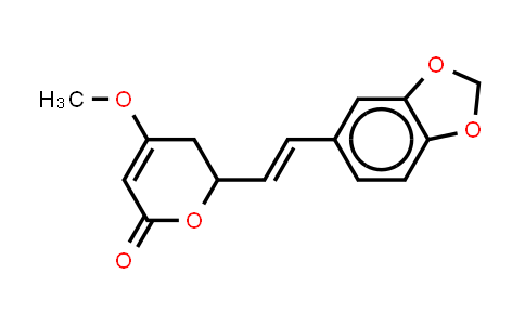
| Chemical Name |
Methysticin |
| CAS Number |
20697-20-5 |
| MDL Number |
MFCD18430287 |
| Molecular Formula |
C15H14O5 |
| Molecular Weight |
274.27 |
| Synonyms |
DL-Methysticin;(±)-Methystici |
Introduction of 20697-20-5 :
Methysticin is a major kavalactone in kava extract to induce CYP1A1. IC50 & Target: CYP1A1[1] In Vitro: Methysticin triggers the most profound inducing effect on CYP1A1. Consistent with the experimental results, in silico molecular docking studies based on the aryl hydrocarbon receptor (AhR)-ligand binding domain homology model also reveals favorable binding to AhR for Methysticin compared with the remaining kavalactones. Additionally, results from a luciferase gene reporter assay suggested that kava extract, Methysticin is able to activate the AhR signaling pathway. Kava extract induces the expression of CYP1A1 via an AhR-dependent mechanism and that Methysticin contributes to CYP1A1 induction. The induction of CYP1A1 indicates a potential interaction between kava or kavalactones and CYP1A1-mediated chemical carcinogenesis. The MTS cell viability assay is used to determine the effects of kava extract and kavalactones on cell viability in mouse hepatic cells. Hepa1c1c7 cells are treated with various concentrations of kava extract (0-50 µg/mL) and six kavalactones (0-100 µM) for 24 h. The results indicate that kava extract at concentrations up to 50 µg/mL and kavalactones up to 100 µM do not induce cell death. For the following studies, kava extract at 0.78-6.25 µg/mL and kavalactones at 0.78-25 µM, concentrations that cause no damage to cells, are used[1]. In Vivo: The kavalactone Methysticin (6 mg/kg) is administered once a week for a period of 6 months to 6 month old transgenic APP/Psen1 mice by oral gavage. Methysticin treatment activates the Nrf2 pathway in the hippocampus and cortex of mice. The Aβ deposition in brains of Methysticin-treated APP/Psen1 mice is not altered compared to untreated mice. However, Methysticin treatment significantly reduces microgliosis, astrogliosis and secretion of the pro-inflammatory cytokines TNF-α and IL-17A. Methysticin treatment results in a significant activation of the Nrf2/ARE pathway in hippocampus and the cortex but not in the midbrain and cerebellum of ARE-luciferase reporter gene mice. Methysticin treatment significantly increases the expression of both genes compared to untreated animals[2].
| Purity |
NLT 98% |
| Storage |
at 20ºC 2 years |
*The above information is for reference only.
